Human hip bone


Human hip bone
Published 2018-04-24T11:09:36+00:00
University: Pavia University
Faculty: Engineering
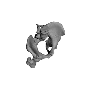
Hello, I am a biomedical engineering student of the University of Pavia in Italy and I wanted to realize in 3D a part of our body, in this case the hip bone of a patient.
To perform this work I needed a CT scan and a program that could read the respective DICOM files, after which I isolated the affected organ, in this case the hip bone, and I converted it into an STL file that could be printed.
These kinds of approaches to medicine is very useful for doctors, since they manage to approach an intervention better. It is much better to have the organ at 1:1 scale in your hand than to see images in two dimensions. Our hospital in Pavia already uses this technology.
Note:
The hip bone is at 1: 2 scale, it can be modified so as to print it to any size.


Rafts: No
Supports: YES
Resolution: 0.1, 0.15
Infill: 15%
| Date published | 24/04/2018 |
| Material Quantity | ~200g |
| Dimensions | 82mm X 146mm X 124mm |
| Technology | FDM |
| Complexity | Easy |